{Polascin.Net}     {SK.polascin.net}     {Polascin.Sk}     {Books}     {polascin.org}     {nephro.site}     {nephrosite.polascin.net}  

Day:     Year:     Week:     Today is 
()
Lubomir Polascin
Personal WebSite
Lubomir Polascin's Face in Photograph
53
(53.784)
51.271       76.807       75.557       49.867
Ordinácia

Bio

Lubomir Polascin
Pronounced like Ljubomeer Polaschtcheen

Contacts E-Mail:   lubomir@polascin.net
Web:     polascin.net  | sk.polascin.net  | polascin.sk
I appreciate it if you could refrain from contacting me by phone if you are not within my personal or work circle. Thank you.
LinkedIn Logo


I am a medical doctor who graduated in General Medicine (1995) and specialized in Nephrology (2009). I am attested in Internal Medicine (1998) and certified in Abdominal Ultrasonic Imaging in Adults (2009). My areas of expertise include Dialysis, Blood Purification Therapies, Vascular Access Ultrasonography for Dialysis, Extra-corporeal and Intra-corporeal Elimination Therapies, and Renal Replacement Therapies, such as Hemodialysis, Hemodiafiltration, Hemofiltration, Selective Absorption and Adsorption Treatments, Membrane Plasma Separation - Plasmapheresis, Peritoneal Dialysis, and Kidney Transplant. Additionally, I have experience in teaching and lecturing on these fields, as well as Internal Medicine, Diabetology, and associated fields of medicine.
Translator specialized in English-Slovak and Slovak-English medical translation and software localization.
Information technology, personal and other computers, computer science, algorithms, programming, coding, HTML, PHP, CSS, JavaScript, SQL, web design, creation of websites and applications, Python, data analysis, database systems, language transformers, information technology, internet and communication technology, office and database application software, operating systems, UNIX, Linux, free and open-source software (FOSS), and artificial intelligence and its applications.
Ever since I was a child, I have been an avid reader of books from a diverse range of genres. From science-fiction to fantasy, historical to educational, biographical to personal growth, and even books on alternative perspectives of our reality, perception, and consciousness. My interests extend beyond books, as I am passionate about foreign languages, travel, and the study of language as a phenomenon. I also spend my time studying philosophical works and occasionally indulge in writing and reading poetry. Reading has been an integral part of my life and has broadened my horizons, expanded my imagination, and helped me gain a better understanding of the world around me.

Lubomir Polascin's "About the Author" on Amazon Author Central
23andme     23andme
Renal Picture Banner Renal Picture Banner Renal Picture Banner Renal Picture Banner Renal Picture Banner Renal Picture Banner Renal Picture Banner Renal Picture Banner Renal Picture Banner Renal Picture Banner Renal Picture Banner Renal Picture Banner Renal Picture Banner Renal Picture Banner Renal Picture Banner Renal Picture Banner Renal Picture Banner Renal Picture Banner Renal Picture Banner Renal Picture Banner
Visit NephroSite!
NephroSite
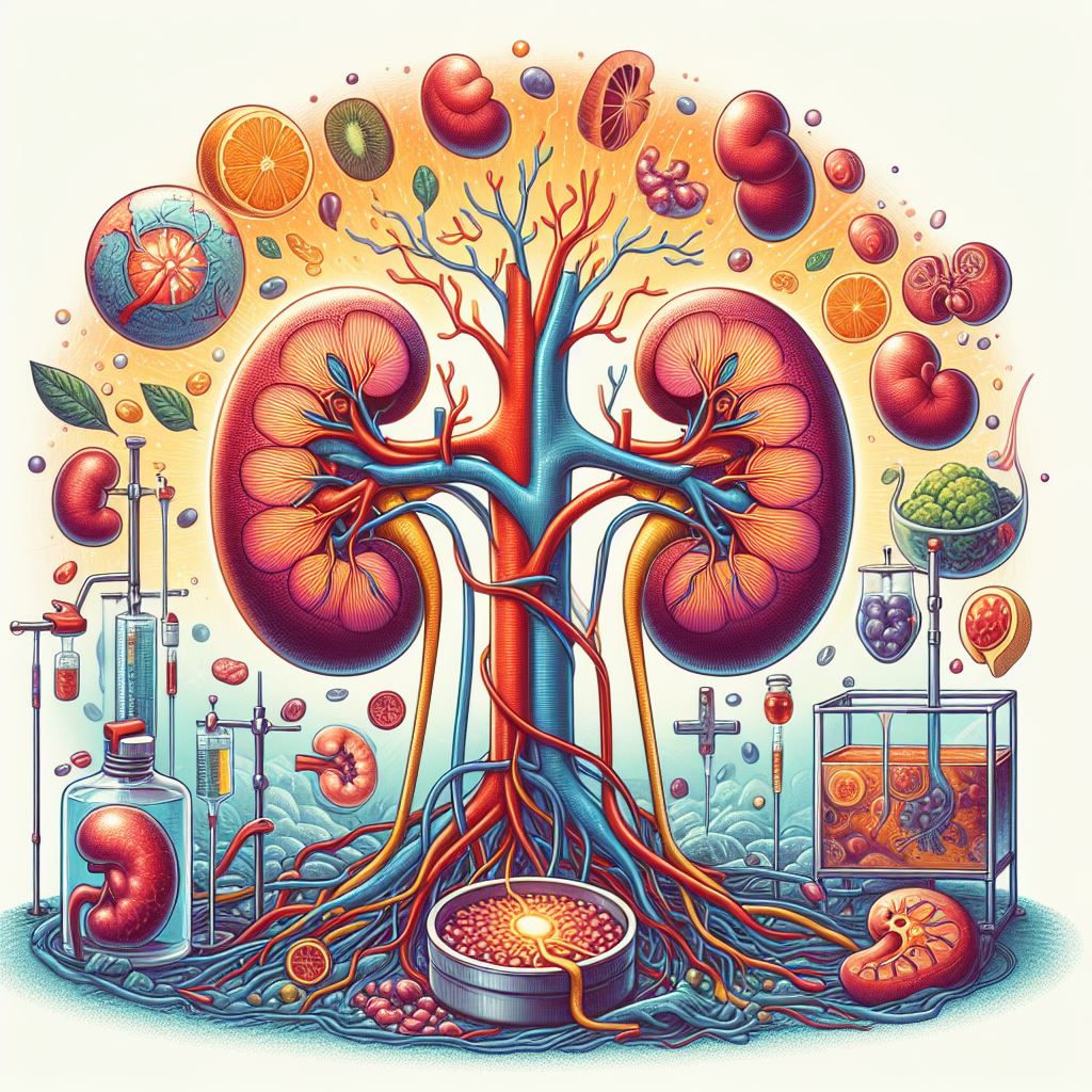
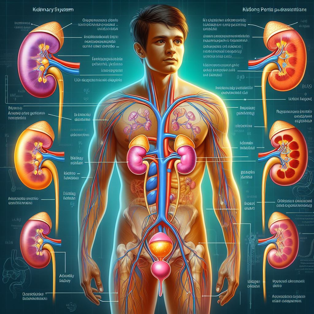
Nephrology Ilustration

Nephrology

Nephrology is an essential medical specialty that deals with the kidneys, which are crucial organs responsible for various vital functions in the human body. The kidneys perform a wide range of tasks, including keeping fluid balance, filtering waste products and excess substances from the bloodstream, regulating blood pressure, and producing hormones involved in red blood cell production and bone health.

As experts in kidney health, nephrologists oversee diagnosing, treating, and managing an array of kidney-related conditions and disorders. These include:

  1. Chronic kidney disease (CKD): This progressive condition occurs when the kidneys lose their ability to function effectively over an extended period. CKD can result from several factors, such as diabetes, hypertension, autoimmune diseases, or genetic conditions. As the disease progresses, patients may need dialysis or a kidney transplant.
  2. Acute kidney injury (AKI): Unlike CKD, AKI is a sudden and temporary loss of kidney function. It can occur due to factors like severe infections, toxic substances, dehydration, or reduced blood flow to the kidneys. Timely intervention is essential to prevent permanent damage or the progression to chronic kidney disease.
  3. Glomerular diseases, Glomerulonephritis (GN): Glomeruli, the kidney's tiny filtering units, can be affected by various diseases, such as glomerulonephritis or diabetic nephropathy. These conditions can cause inflammation, scarring, and damage to the kidneys, impairing their ability to filter waste and keep fluid balance.
  4. Kidney stones: These are solid deposits of minerals and salts that form within the kidneys. They can cause intense pain, infection, and obstruct urine flow. Nephrologists can help manage kidney stones through medication, lifestyle changes, or minimally invasive procedures.
  5. Tubular disorders: The renal tubules manage reabsorbing essential substances and secreting waste products. Conditions like renal tubular acidosis or Fanconi syndrome affect the tubules' proper functioning, leading to electrolyte imbalances and other complications.
  6. Renal artery stenosis: This condition occurs when the renal arteries, which supply blood to the kidneys, become narrowed due to plaque build-up or other factors. Reduced blood flow can impair kidney function and contribute to high blood pressure.
  7. Polycystic kidney disease (PKD): A genetic disorder characterized by the growth of numerous cysts in the kidneys, leading to reduced kidney function and eventual kidney failure.
  8. Electrolyte imbalances: Abnormal levels of essential electrolytes in the body, such as sodium, potassium, and calcium, which can be caused by kidney dysfunction.
  9. Hypertension (high blood pressure): Elevated blood pressure that can damage the blood vessels in the kidneys and impair kidney function.
  10. Kidney infections: Infections like pyelonephritis result from bacteria entering the kidneys, usually from the urinary tract. Symptoms may include fever, back pain, and frequent urination. Antibiotics and other treatments are essential to prevent complications.

Nephrologists employ various diagnostic tools and tests to evaluate kidney function, including blood tests, urine tests, imaging studies (e.g., ultrasound, CT scans), and kidney biopsies. They supply tailored treatment plans for each patient, which may involve medications, dietary modifications, lifestyle changes, or medical procedures like dialysis and kidney transplantation.

In summary, nephrology is a vital medical specialty that deals with the diagnosis, treatment, and management of kidney-related conditions. Nephrologists play a crucial role in supporting kidney health and helping patients manage their conditions to improve their overall quality of life.

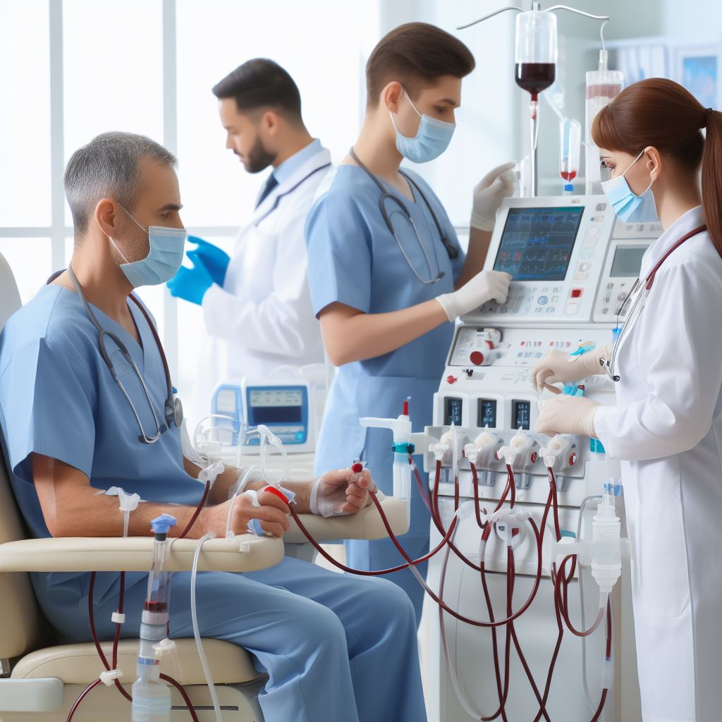
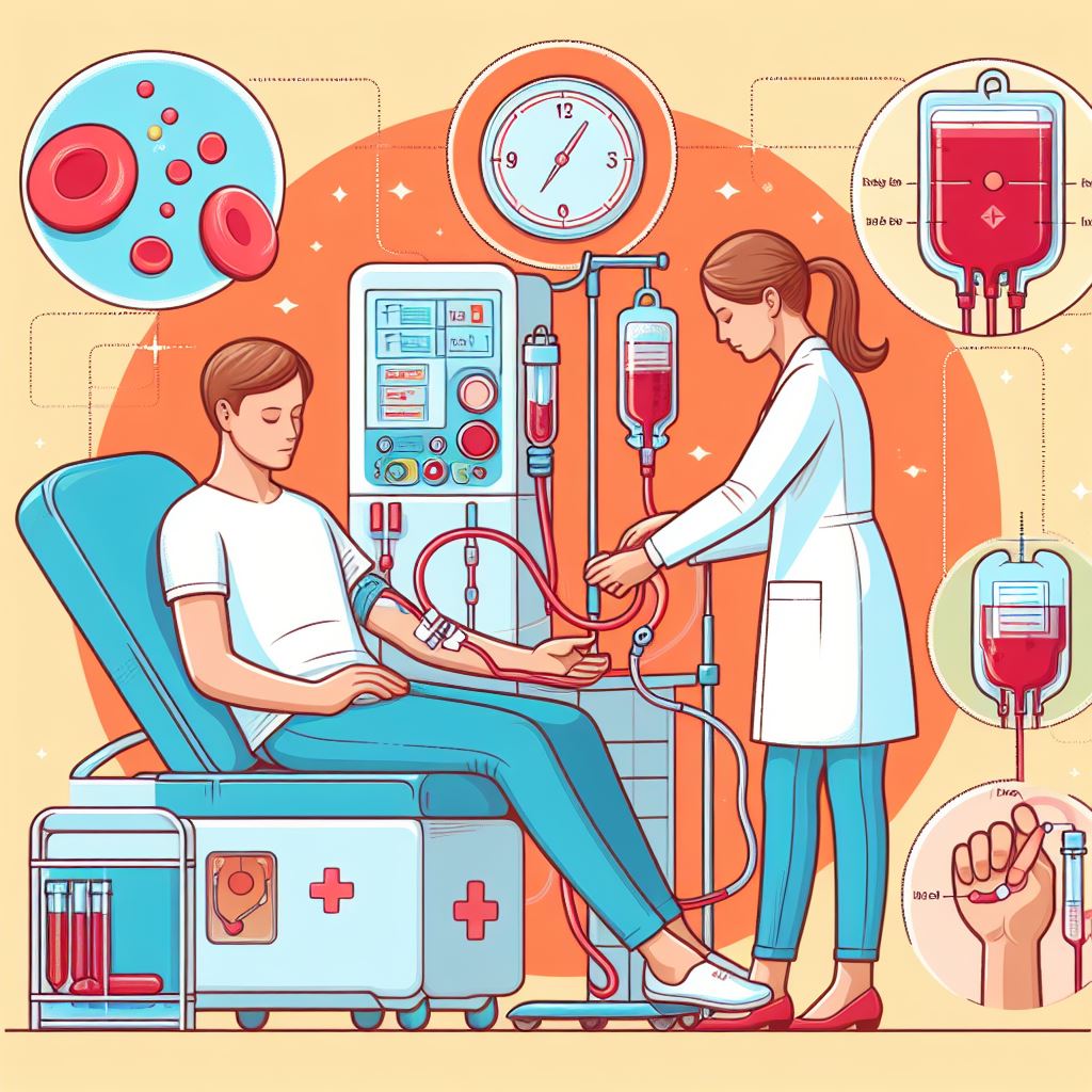
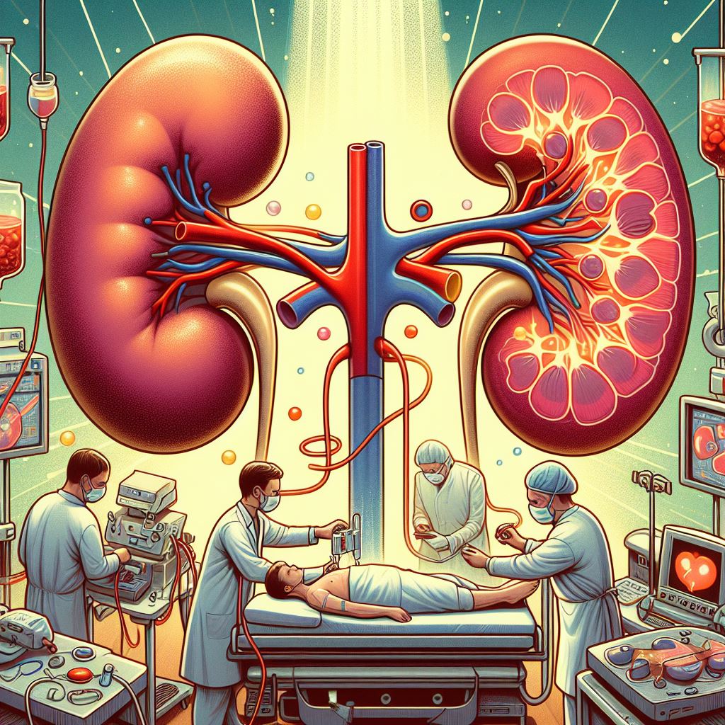
Dialysis Ilustration

Dialysis

Dialysis is a medical treatment that performs some of the kidneys' essential functions when they can no longer function effectively on their own. The procedure helps to filter waste products, excess fluids, and electrolytes from the bloodstream, serving as a lifesaving intervention for individuals with end-stage renal disease (ESRD) [end-stage kidney disease (ESKD)] or acute kidney injury (AKI).

Principles of Dialysis. Dialysis relies on the principles of diffusion and ultrafiltration to separate waste products and excess fluids from the blood. Diffusion refers to the passive movement of solutes across a semipermeable membrane, while ultrafiltration involves the application of pressure to force fluid through the membrane. By using these principles, dialysis helps to keep proper blood chemistry and fluid balance in the body.

Types of Dialysis. There are two primary types of dialysis:

  1. Haemodialysis: This is the most ordinary form of dialysis, in which blood is filtered through an artificial kidney machine, known as a dialyzer. The dialyzer holds a semipermeable membrane that separates waste products and excess fluids from the blood, which are then discarded, and the cleaned blood is returned to the body.
  2. Peritoneal dialysis: This type of dialysis uses the body's peritoneal membrane, which lines the abdominal cavity, as a natural semipermeable filter. A sterile dialysis solution is introduced into the abdomen through a catheter, and waste products and excess fluids pass from the blood into the solution through the peritoneal membrane. The solution is then drained and replaced with fresh solution, typically several times per day.

Indications for Dialysis. Dialysis is typically written down for individuals with:

  1. End-stage renal disease (ESRD): When chronic kidney disease progresses to a point where the kidneys can no longer sustain life, dialysis becomes necessary to replace lost kidney function.
  2. Acute kidney injury (AKI): Dialysis may be needed temporarily to support patients with sudden kidney failure until their kidney function recovers.
  3. Severe electrolyte imbalances or fluid overload: Dialysis can help correct life-threatening imbalances or manage fluid overload when other treatments are ineffective.

Benefits of Dialysis. Dialysis offers several essential benefits for patients with kidney failure, including:

  1. Removal of waste products and toxins: Dialysis helps support proper blood chemistry by filtering harmful substances that would otherwise accumulate in the body.
  2. Fluid and electrolyte balance: Dialysis helps regulate fluid levels and correct electrolyte imbalances, reducing the risk of complications such as oedema or heart problems.
  3. Improved quality of life: By managing the symptoms and complications of kidney failure, dialysis can enhance patients' overall well-being and enable them to lead more active lives.

Risks and Complications. While dialysis is a lifesaving intervention, it carries potential risks and complications, including:

  1. Infection: Both haemodialysis and peritoneal dialysis are associated with an increased risk of infection, particularly at the site of vascular access or peritoneal catheter.
  2. Low blood pressure: Dialysis may cause a drop in blood pressure, leading to dizziness, fainting, or in severe cases, shock.
  3. Anaemia: Dialysis can contribute to anaemia, as the process may cause some blood loss and affect the production of red blood cells.
  4. Muscle cramps: Rapid fluid removal during dialysis can cause muscle cramps, particularly in the legs.
  5. Peritonitis: Peritoneal dialysis carries the risk of peritonitis, an inflammation of the peritoneal membrane caused by bacterial infection.
  6. Disequilibrium syndrome: A rare complication of haemodialysis, disequilibrium syndrome occurs when the blood is cleaned too quickly, leading to symptoms like nausea, vomiting, headache, and, in severe cases, seizures or coma.

Impact on Patients' Quality of Life. Dialysis can significantly affect patients' quality of life, as it requires a substantial time commitment and lifestyle adjustments. Factors that affect patients on dialysis include:

  • Time commitment: Haemodialysis typically needs three to four sessions per week, each lasting three to five hours, while peritoneal dialysis involves multiple exchanges per day or overnight treatments with a machine. This can limit patients' ability to work, travel, or engage in leisure activities.
  • Dietary restrictions: Patients on dialysis often need to follow a strict diet to manage fluid intake, potassium, phosphorus, and sodium levels, which can be challenging to adhere to.
  • Emotional and psychological effects: The demands of dialysis and the reality of living with a chronic illness can lead to feelings of depression, anxiety, or isolation for some patients.
  • Financial burden: Dialysis treatments can be costly, and the associated expenses may place a significant financial strain on patients and their families.

Despite these challenges, many patients on dialysis can support a fulfilling and active life with proper support, education, and self-care. By working closely with their healthcare team, patients can develop strategies to manage the demands of dialysis and adapt to their new lifestyle.

Dialysis is a critical treatment choice for individuals with kidney failure, supplying lifesaving support when the kidneys can no longer function effectively. The procedure offers essential benefits, such as waste removal, fluid balance, and improved quality of life, but also carries potential risks and complications. Understanding the principles, types, indications, and impacts of dialysis is crucial for patients and their families, as it empowers them to make informed decisions about their care and navigate the challenges associated with living with kidney failure.

Haemodialysis Ilustration

Haemodialysis

Haemodialysis is a medical treatment that filters waste products, excess fluids, and electrolytes from the bloodstream when the kidneys can no longer function effectively. It is the most usual form of dialysis and serves as a lifesaving intervention for individuals with end-stage renal disease (ESRD) or acute kidney injury (AKI).

Principles of Haemodialysis. Haemodialysis relies on the principles of diffusion, adsorption, convection, and ultrafiltration to separate waste products and excess fluids from the blood. During the procedure, blood is circulated through an artificial kidney machine, known as a dialyzer, which holds a semipermeable membrane. Waste products, excess fluids, and electrolytes pass through the membrane and into a dialysis solution, which is then discarded. The cleaned blood is returned to the body, helping keep proper blood chemistry and fluid balance.

Components of Haemodialysis. There are three main components of haemodialysis:

  1. Dialyzer: The artificial kidney machine consists of a semipermeable membrane that filters the blood. The dialyzer's membrane is usually made from synthetic materials, such as cellulose or synthetic polymers, which supply best filtration properties.
  2. Dialysis solution: Also known as dialysate, this sterile solution holds water and electrolytes that help ease the removal of waste products and the balance of electrolytes in the blood.
  3. Vascular access: This is the point of entry for the blood to be removed from the body, filtered through the dialyzer, and returned to the body. There are three primary types of vascular access: arteriovenous (AV) fistula, AV graft, and central venous catheter.

The Haemodialysis Procedure. The haemodialysis procedure typically involves the following steps:

  1. Preparation: The patient's weight, blood pressure, and temperature are checked before starting the treatment. The dialysis machine is set up with the proper dialyzer and dialysis solution.
  2. Vascular access: The patient's vascular access site (AV fistula, AV graft, or central venous catheter) is cleaned and prepared for needle insertion. For patients with an AV fistula or graft, two needles are inserted - one to draw blood out and the other to return the cleaned blood.
  3. Blood circulation: The dialysis machine pumps the blood through the dialyzer at a controlled rate. As the blood passes through the semipermeable membrane, waste products, excess fluids, and electrolytes are removed and discarded.
  4. Monitoring: Healthcare professionals closely watch the patient's vital signs and the dialysis machine settings throughout the treatment to ensure safety and effectiveness.
  5. Completion: After the prescribed treatment time, which usually lasts three to five hours, the blood flow is stopped, the needles are removed, and the vascular access site is cleaned and dressed.
  6. Post-treatment assessment: The patient's weight and blood pressure are checked again, and any concerns or complications are addressed before they leave the dialysis centre.

Benefits of Haemodialysis. Haemodialysis offers several essential benefits for patients with kidney failure, including:

  1. Removal of waste products and toxins: Haemodialysis helps keep proper blood chemistry by filtering harmful substances that would otherwise accumulate in the body.
  2. Fluid and electrolyte balance: Haemodialysis helps regulate fluid levels and correct electrolyte imbalances, reducing the risk of complications such as oedema or heart problems.
  3. Improved quality of life: By managing the symptoms and complications of kidney failure, haemodialysis can enhance patients' overall well-being and enable them to lead more active lives.

Risks and Complications of Haemodialysis. While haemodialysis is a lifesaving intervention, it carries potential risks and complications, including:

  1. Infection: Infections can occur at the site of vascular access or in the bloodstream, particularly if proper hygiene practices are not followed.
  2. Low blood pressure: Haemodialysis may cause a drop in blood pressure, leading to dizziness, fainting, or in severe cases, shock.
  3. Anaemia: Haemodialysis can contribute to anaemia, as the process may cause some blood loss and affect the production of red blood cells.
  4. Muscle cramps: Rapid fluid removal during haemodialysis can cause muscle cramps, particularly in the legs.
  5. Disequilibrium syndrome: A rare complication of haemodialysis, disequilibrium syndrome occurs when the blood is cleaned too quickly, leading to symptoms like nausea, vomiting, headache, and, in severe cases, seizures or coma.

Impact on Patients' Quality of Life. Haemodialysis can significantly affect patients' quality of life, as it requires a substantial time commitment and lifestyle adjustments. Factors that affect patients on haemodialysis include:

  • Time commitment: Haemodialysis typically needs three to four sessions per week, each lasting three to five hours. This can limit patients' ability to work, travel, or engage in leisure activities.
  • Dietary restrictions: Patients on haemodialysis often need to follow a strict diet to manage fluid intake, potassium, phosphorus, and sodium levels, which can be challenging to adhere to.
  • Emotional and psychological effects: The demands of haemodialysis and the reality of living with a chronic illness can lead to feelings of depression, anxiety, or isolation for some patients.
  • Financial burden: Haemodialysis treatments can be costly, and the associated expenses may place a significant financial strain on patients and their families.

Despite these challenges, many patients on haemodialysis can support a fulfilling and active life with proper support, education, and self-care. By working closely with their healthcare team, patients can develop strategies to manage the demands of haemodialysis and adapt to their new lifestyle.

Haemodialysis is a crucial treatment choice for individuals with kidney failure, supplying lifesaving support when the kidneys can no longer function effectively. The procedure offers essential benefits, such as waste removal, fluid balance, and improved quality of life, but also carries potential risks and complications. Understanding the principles, components, procedure, and impacts of haemodialysis is vital for patients and their families, as it empowers them to make informed decisions about their care and navigate the challenges associated with living with kidney failure.

Lubomir Polascin, MD at BORY HOSPITAL, Bratislava
Peritoneal Dialysis Ilustration

Peritoneal Dialysis

Peritoneal dialysis (PD) is a form of dialysis that uses the body's peritoneal membrane as a natural filter to remove waste products, excess fluids, and electrolytes from the bloodstream when the kidneys can no longer function effectively. PD is a home-based treatment choice for individuals with end-stage renal disease (ESRD) or acute kidney injury (AKI) and offers an alternative to haemodialysis.

Principles of Peritoneal Dialysis. Peritoneal dialysis relies on the principles of diffusion and osmosis to separate waste products and excess fluids from the blood. The peritoneal membrane, which lines the abdominal cavity, acts as a natural semipermeable filter. A sterile dialysis solution, called dialysate, is introduced into the abdomen through a catheter, and waste products and excess fluids pass from the blood into the dialysate through the peritoneal membrane. The used dialysate is then drained and replaced with fresh solution.

Types of Peritoneal Dialysis. There are two primary types of peritoneal dialysis:

  1. Continuous Ambulatory Peritoneal Dialysis (CAPD): CAPD is a manual form of PD that involves performing several exchanges of dialysate throughout the day, usually four to five times. Each exchange involves draining the used dialysate, filling the abdomen with fresh dialysate, and allowing the solution to dwell for a specified period (usually 4-6 hours) before repeating the process.
  2. Automated Peritoneal Dialysis (APD): APD, also known as Continuous Cycling Peritoneal Dialysis (CCPD), uses a machine called a cycler to perform the exchanges. The cycler automatically fills and drains the abdomen with dialysate over a specified period, typically during night-time sleep. This allows the patient to have a more flexible daytime schedule without the need for manual exchanges.

The Peritoneal Dialysis Procedure. The peritoneal dialysis procedure typically involves the following steps:

  1. Catheter placement: A soft, flexible catheter is surgically inserted into the patient's abdomen, usually a few weeks before starting PD to allow healing. The catheter serves as a portal for introducing and draining the dialysate.
  2. Preparing the dialysis solution: The patient prepares the sterile dialysate by warming it to body temperature and connecting it to the transfer set, a tubing system that links the dialysate bag to the catheter.
  3. Performing the exchange: For CAPD, the patient manually drains the used dialysate from the abdomen, fills the abdomen with fresh dialysate, and allows the solution to dwell for a specified period before repeating the process. For APD, the cycler automatically manages the filling, dwelling, and draining processes during night-time sleep.
  4. Monitoring and follow-up: Regular check-ups with healthcare professionals are essential for evaluating the effectiveness of PD, assessing the catheter site for signs of infection, and adjusting the dialysis prescription as needed.

Benefits of Peritoneal Dialysis. Peritoneal dialysis offers several advantages for patients with kidney failure, including:

  1. Home-based treatment: PD allows patients to perform dialysis at home, supplying greater flexibility and independence compared to in-centre haemodialysis.
  2. Fewer dietary restrictions: PD patients typically have fewer dietary restrictions than those on haemodialysis, as the continuous nature of the treatment helps keep more stable blood chemistry.
  3. Improved quality of life: PD often results in fewer fluctuations in blood chemistry, which can lead to a better overall sense of well-being and a more stable energy level.
  4. Better preservation of residual kidney function: PD is generally gentler on the remaining kidney function compared to haemodialysis, which may slow the progression of kidney disease.
  5. No needles: Unlike haemodialysis, PD does not need needle insertions for vascular access, which can be a significant advantage for patients with needle phobia or difficult vascular access.

Risks and Complications of Peritoneal Dialysis. While peritoneal dialysis offers many benefits, it is not without potential risks and complications, including:

  1. Peritonitis: Infection of the peritoneal membrane, known as peritonitis, is the most common complication of PD. It is often caused by bacteria introduced during exchanges or from an infected catheter site. Prompt treatment with antibiotics is essential to prevent profound consequences.
  2. Catheter-related issues: Infections or mechanical problems with the catheter, such as blockage or dislodgement, can occur, requiring medical intervention.
  3. Hernias: The increased pressure within the abdomen from the dialysate may increase the risk of developing a hernia, particularly in patients with pre-existing weak spots in the abdominal wall.
  4. Fluid and electrolyte imbalances: While PD generally maintain stable blood chemistry, it may sometimes lead to imbalances in fluid or electrolyte levels that require adjustments to the dialysis prescription.
  5. Protein loss: The peritoneal membrane may allow some protein to leak into the dialysate, which can result in a decrease in blood protein levels over time. In some cases, this may require dietary or medication adjustments.

Impact on Patients' Quality of Life. Peritoneal dialysis can have both positive and negative effects on patients' quality of life:

  • Independence and flexibility: PD offer greater independence and flexibility compared to in-centre haemodialysis, allowing patients to perform the treatment at home and support a more normal daily routine.
  • Time commitment: While PD does not need lengthy in-centre treatments, it still demands regular exchanges throughout the day (for CAPD) or nightly treatments (for APD), which can be time-consuming.
  • Self-management: PD requires patients to take an active role in their treatment, including monitoring vital signs, performing exchanges, and keeping a sterile environment. This can be empowering for some patients, while others may find it burdensome or stressful.
  • Emotional and psychological effects: As with any chronic illness, living with kidney failure and undergoing dialysis can lead to feelings of depression, anxiety, or isolation for some patients.

Peritoneal dialysis is a valuable treatment choice for individuals with kidney failure, offering an alternative to haemodialysis that allows for greater independence, flexibility, and a more stable blood chemistry. While PD carries potential risks and complications, many patients find that the benefits of home-based treatment and the opportunity for self-management contribute to an improved quality of life. Understanding the principles, types, procedure, benefits, and impacts of peritoneal dialysis is essential for patients and their families, as it empowers them to make informed decisions about their care and navigate the challenges associated with living with kidney failure.
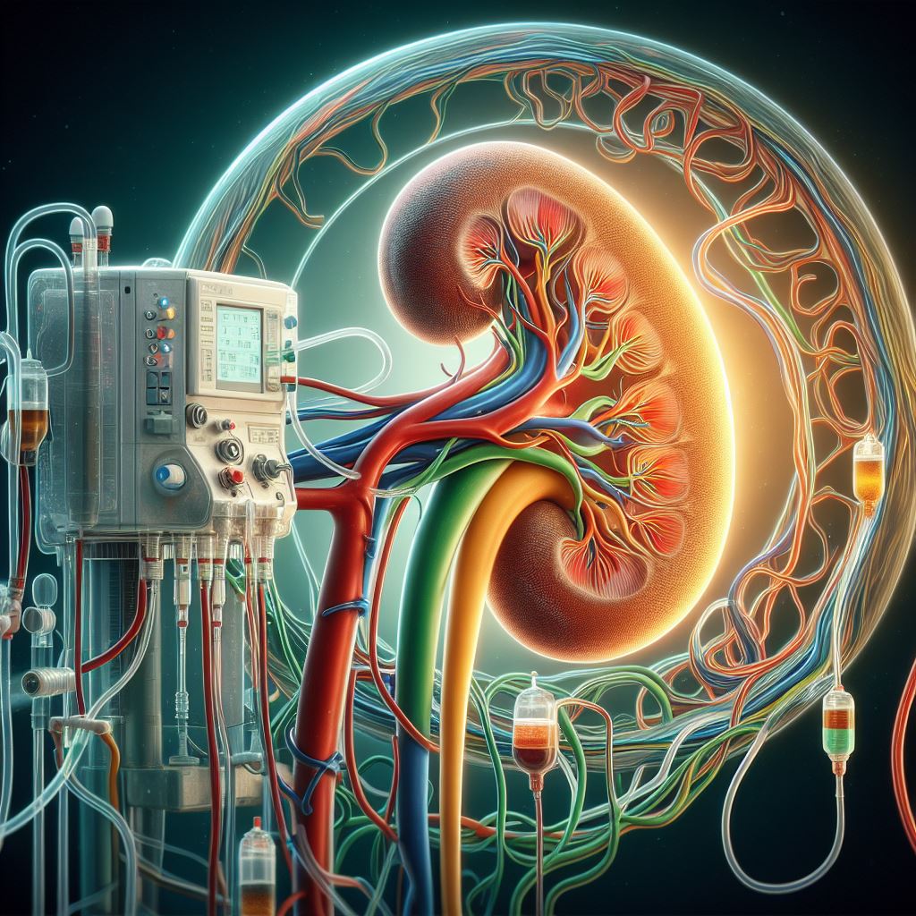
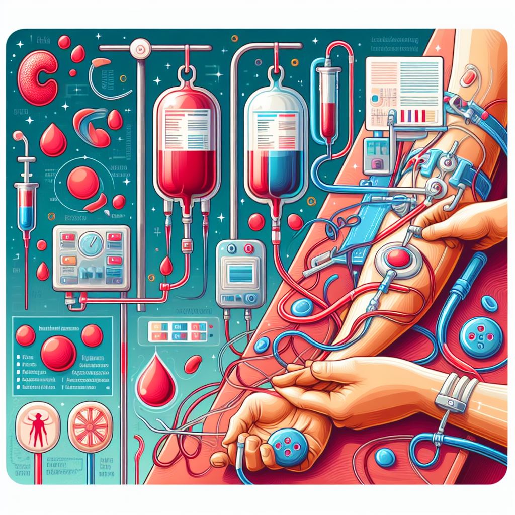
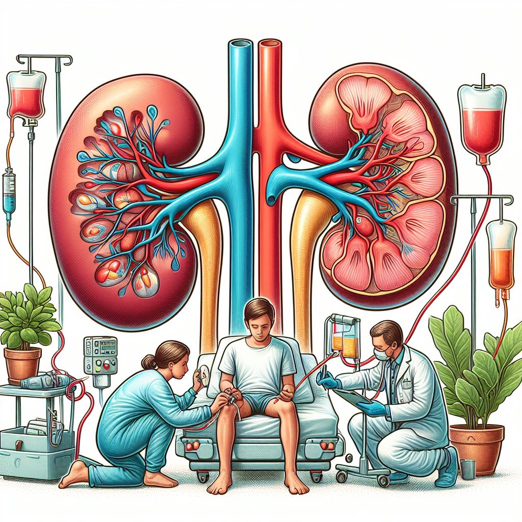
Blood Purification Ilustration

Blood Purification

Blood purification is a general term for various medical therapies and techniques designed to remove harmful substances, toxins, and waste products from the bloodstream. These treatments are often employed when the body's natural detoxification processes, such as the liver and kidneys, are compromised or overwhelmed. Blood purification therapies are primarily used for patients with kidney failure, liver failure, or severe poisoning.

  1. Haemodialysis
    Haemodialysis is the most ordinary form of blood purification used to treat patients with kidney failure. In haemodialysis, blood circulates through an external filter called a dialyzer, where waste products, excess fluids, and electrolytes are removed through a semipermeable membrane. The cleansed blood is then returned to the patient's body. Haemodialysis is typically performed in specialized dialysis centres and requires sessions lasting several hours, multiple times per week.
  2. Hemofiltration
    Hemofiltration is a blood purification technique like haemodialysis but primarily focuses on removing excess fluid and larger molecules, such as inflammatory mediators and cytokines. This therapy utilizes a filtration membrane with larger pores, allowing more significant substances to be removed from the blood. Hemofiltration is often used in the management of acute kidney injury, sepsis, or in combination with haemodialysis.
  3. Hemoperfusion
    Hemoperfusion is a blood purification technique specifically designed to remove toxic substances, such as drugs or poisons, from the bloodstream. In this procedure, blood is passed through a column containing an adsorbent material, such as activated charcoal or a specific resin, which binds to the toxic substance and removes it from the blood. Hemoperfusion is commonly used in cases of drug overdose, poisoning, or acute liver failure.
  4. Plasmapheresis
    Plasmapheresis, also known as therapeutic plasma exchange (TPE), is a blood purification technique that involves separating blood components and selectively removing harmful substances from the plasma. In this procedure, blood is removed from the patient's body and passed through a machine that separates the plasma from the blood cells. The plasma, which contains the harmful substances, is discarded, and replaced with a plasma substitute or donor plasma. The cleansed blood components are then returned to the patient's body. Plasmapheresis is used in the treatment of various autoimmune and neurological disorders, such as myasthenia gravis, Guillain-Barré syndrome, and thrombotic thrombocytopenic purpura.
  5. Immunoadsorption
    Immunoadsorption is a blood purification technique that selectively removes specific immune-related substances, such as autoantibodies, immune complexes, or cytokines, from the blood. This procedure is performed by circulating the blood through a column containing an adsorbent material with a high affinity for the target substance. Immunoadsorption is used in the treatment of autoimmune disorders, such as systemic lupus erythematosus, and in some cases of transplantation to reduce the risk of organ rejection.

Blood purification techniques play a crucial role in the management of various medical conditions where the body's natural detoxification processes are compromised or overwhelmed. By understanding the different blood purification methods, such as haemodialysis, hemofiltration, hemoperfusion, plasmapheresis, and immunoadsorption, healthcare professionals can select the most appropriate therapy based on the patient's specific needs and medical condition. These treatments can help improve patient outcomes and enhance their quality of life.

Renal Transplantation Ilustration

Renal Transplantation

Renal transplantation, also known as kidney transplantation, is a surgical procedure in which a healthy kidney from a living or deceased donor is transplanted into a patient with end-stage renal disease (ESRD) or acute kidney injury (AKI). Kidney transplantation is often considered the best long-term treatment option for kidney failure, as it can supply a higher quality of life and better survival rates compared to dialysis.

Evaluation Process for Renal Transplantation. Before a patient can be considered for a kidney transplant, they must undergo a thorough evaluation process, which typically includes:

  • Medical assessment: A comprehensive examination of the patient's medical history, current health status, and kidney function to decide if they are a suitable candidate for transplantation.
  • Compatibility testing: Blood tests, including blood type and tissue typing, are performed to assess the compatibility between the potential donor and recipient to minimize the risk of organ rejection.
  • Psychosocial assessment: A review of the patient's mental and emotional well-being, as well as their social support network, to ensure they are prepared for the demands of post-transplant care.
  • Financial considerations: An evaluation of the patient's financial resources and insurance coverage, as transplantation and post-transplant care can be expensive.

Donor Types for Renal Transplantation. There are two primary types of kidney donors:

  1. Living donors: Living donors can be related (e.g., siblings, parents, or children) or unrelated (e.g., friends, spouses, or altruistic strangers) to the recipient. Living donor transplantation offers several advantages, such as better compatibility, shorter waiting times, and improved kidney function and survival rates compared to deceased donor transplantation.
  2. Deceased donors: Deceased donors are individuals who have suffered brain death or circulatory death and have agreed to donate their organs. Deceased donor kidneys are distributed to recipients based on several factors, such as blood type, tissue match, waiting time, and medical urgency.

The Renal Transplantation Procedure. The kidney transplantation procedure typically involves the following steps:

  1. Anaesthesia: The recipient is placed under general anaesthesia, making them unconscious and pain-free during the surgery.
  2. Incision: A surgical incision is made in the lower abdomen, usually on the side of the non-functioning kidney.
  3. Placement of the donor kidney: The donor kidney is placed in the recipient's lower abdomen and connected to the recipient's blood vessels and ureter. The original kidneys are usually left in place unless they are causing complications.
  4. Closure: Once the connections are secure and blood flow is set up, the incision is closed with sutures or staples.

Post-Transplant Care and Monitoring:. After the transplantation, the recipient will require close monitoring and lifelong care to ensure the success of the transplant and support their overall health:

  1. Immunosuppressive medications: The recipient will need to take immunosuppressive medications to prevent their immune system from attacking the transplanted kidney (rejection). These medications must be taken consistently and adjusted periodically based on the patient's response and kidney function.
  2. Regular check-ups: The recipient will need regular check-ups with their transplant team to watch kidney function, assess for signs of rejection, and adjust medications as needed.
  3. Lifestyle adjustments: The recipient will need to support a healthy lifestyle, including a balanced diet, regular exercise, and proper hydration to support the transplanted kidney's function and overall health.

Benefits of Renal Transplantation. Kidney transplantation offers several significant benefits for patients with kidney failure, including:

  1. Improved quality of life: Transplant recipients often experience a higher quality of life compared to dialysis patients, as they have fewer dietary restrictions, increased energy levels, and greater freedom to engage in work, travel, and leisure activities.
  2. Better survival rates: Kidney transplantation generally provides better long-term survival rates than dialysis, particularly when the kidney is from a living donor.
  3. Lower treatment burden: Unlike dialysis, which requires ongoing treatment sessions, a successful kidney transplant can supply long-term relief from the need for dialysis, allowing patients to lead a more normal life.

Risks and Complications of Renal Transplantation. Despite the many benefits, kidney transplantation carries potential risks and complications, including:

  1. Organ rejection: The recipient's immune system may recognize the transplanted kidney as foreign and attack it, leading to rejection. Rejection can be acute (occurring within days or weeks) or chronic (developing over months or years).
  2. Infections: Immunosuppressive medications, which are necessary to prevent organ rejection, can increase the risk of infections, as they suppress the immune system's ability to fight off bacteria, viruses, and fungi.
  3. Side effects of immunosuppressive medications: These medications can have various side effects, including increased risk of certain cancers, high blood pressure, high cholesterol, diabetes, and bone disease.
  4. Surgical complications: As with any surgery, there is a risk of complications such as bleeding, blood clots, or anaesthesia-related issues.

Impact on Patients' Quality of Life. A successful kidney transplant can have a profound impact on the recipient's quality of life:
  • Freedom from dialysis: Transplant recipients no longer need dialysis, which frees up a significant amount of time and allows them to take part in activities that may have been restricted while on dialysis.
  • Increased energy and well-being: With improved kidney function, transplant recipients often report increased energy levels and a better overall sense of well-being.
  • Greater independence: Kidney transplantation enables patients to lead more independent lives, with fewer restrictions on work, travel, and social activities.

Renal transplantation is a life-changing treatment option for individuals with kidney failure, offering an improved quality of life and better survival rates compared to dialysis. The evaluation process, donor types, surgical procedure, post-transplant care, benefits, risks, and impacts on quality of life are all essential aspects to consider for patients and their families as they navigate the challenges associated with living with kidney failure. By working closely with their healthcare team, patients can make informed decisions about their care and pursue the best possible outcomes.

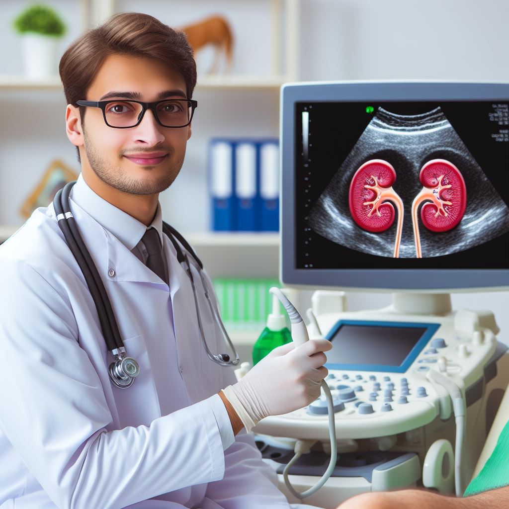
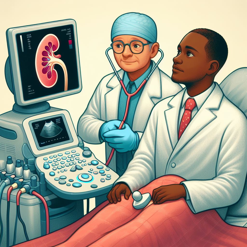
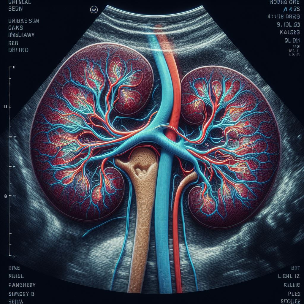
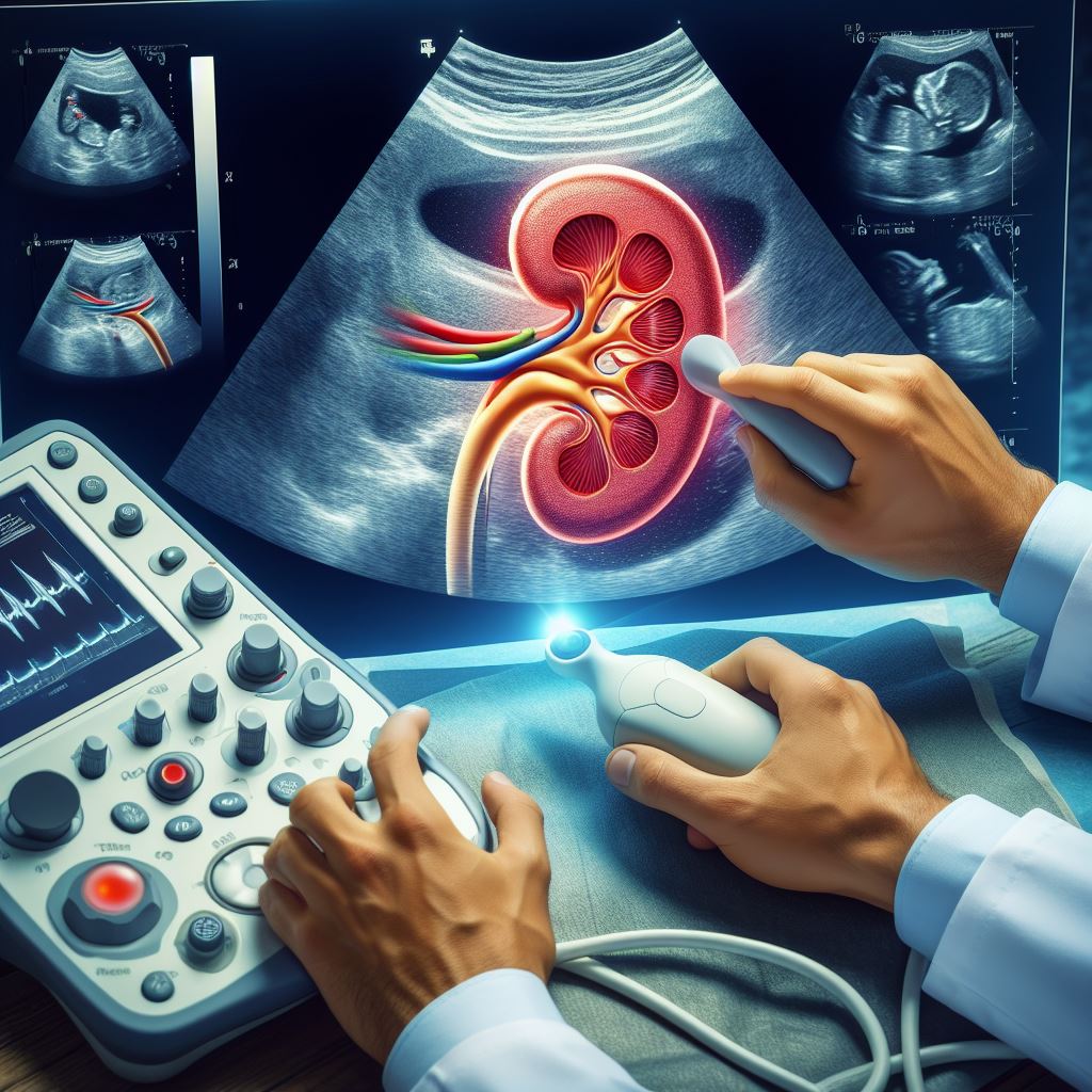
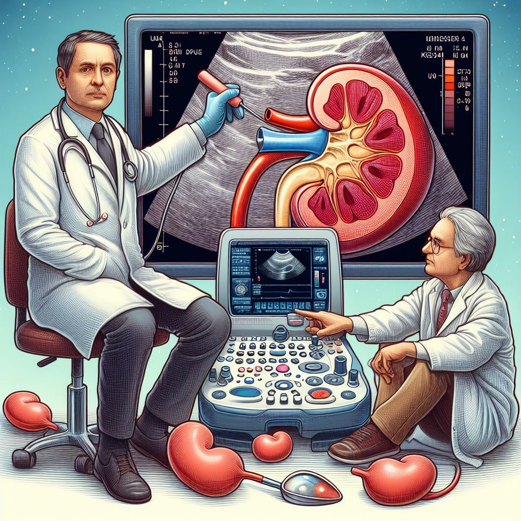
Renal Ultrasound Ilustration

Renal Ultrasound

Kidney ultrasound, also known as renal ultrasound or sonography, is a non-invasive diagnostic imaging technique that uses high-frequency sound waves to create real-time images of the kidneys, ureters, and bladder. Here are the principles, applications, procedure, benefits, and limitations of kidney ultrasound, emphasizing its role in diagnosing and watching various kidney-related conditions.

Principles of Kidney Ultrasound: Ultrasound imaging is based on the principle of echolocation, where high-frequency sound waves are transmitted into the body and then reflected by internal structures. The returning sound waves, or echoes, are converted into electrical signals and processed by a computer to create detailed images of the organs and tissues.

Applications of Kidney Ultrasound: Kidney ultrasound is an essential tool for diagnosing and watching a variety of kidney-related conditions, including:

  1. Kidney stones: Detecting and assessing the size, location, and composition of stones within the kidneys or urinary tract.
  2. Kidney cysts: Identifying and characterizing fluid-filled sacs that may develop in the kidneys.
  3. Tumours and masses: Differentiating between benign and malignant growths in the kidneys.
  4. Hydronephrosis: Evaluating the extent of swelling in the kidneys due to urine backup, often caused by an obstruction in the urinary tract.
  5. Kidney infections: Detecting signs of inflammation or abscess formation in the kidneys.
  6. Congenital abnormalities: Showing structural anomalies in the kidneys or urinary tract present since birth.
  7. Renal artery stenosis: Assessing blood flow to the kidneys and detecting narrowing or blockage in the renal arteries.
  8. Chronic kidney disease: Monitoring kidney size, shape, and parenchymal changes associated with long-term kidney dysfunction.

The Kidney Ultrasound Procedure: The kidney ultrasound procedure is simple, painless, and typically takes 30 minutes to an hour. The patient is usually asked to lie down on their back or side, and a water-based gel is applied to the skin over the kidneys to ensure good contact between the ultrasound probe (transducer) and the body. The sonographer gently moves the probe across the skin, capturing real-time images of the kidneys and surrounding structures. The images are displayed on a monitor and saved for further analysis by a radiologist or nephrologist.

Benefits of Kidney Ultrasound: Kidney ultrasound offers several advantages over other imaging modalities, such as:

  1. Non-invasive: No incisions, injections, or exposure to ionizing radiation are involved.
  2. Painless: The procedure is generally comfortable and well-tolerated by patients.
  3. Real-time imaging: Allows for dynamic assessment of kidney function and blood flow.
  4. Cost-effective: Kidney ultrasound is generally less expensive than other imaging techniques, such as computed tomography (CT) or magnetic resonance imaging (MRI).
  5. Widely accessible: Ultrasound equipment is portable and available in most healthcare settings, from large hospitals to smaller clinics.

Limitations of Kidney Ultrasound: Despite its benefits, kidney ultrasound has some limitations:

  1. Limited resolution: Ultrasound may not supply sufficient detail to visualize small structures or subtle abnormalities.
  2. Obstructed view: Overlying gas in the intestines or obesity can interfere with sound wave transmission, affecting image quality.
  3. Operator-dependent: Image acquisition and interpretation can be affected by the skill and experience of the sonographer or radiologist.

Kidney ultrasound is a valuable diagnostic tool for assessing kidney health and diagnosing a wide range of kidney-related conditions. With its non-invasive nature, real-time imaging capabilities, and cost-effectiveness, it plays a vital role in the management and monitoring of kidney diseases. However, it is essential to consider the limitations of ultrasound and, in some cases, employ complementary imaging techniques such as CT or MRI for a more comprehensive evaluation.

Though kidney ultrasound is an indispensable tool for nephrologists and radiologists, it is crucial to ensure that well-trained and experienced professionals perform and interpret the procedure to keep accuracy and consistency. Overall, kidney ultrasound is an essential part in the diagnosis and management of kidney-related conditions, contributing significantly to improving patient outcomes and supporting kidney health.

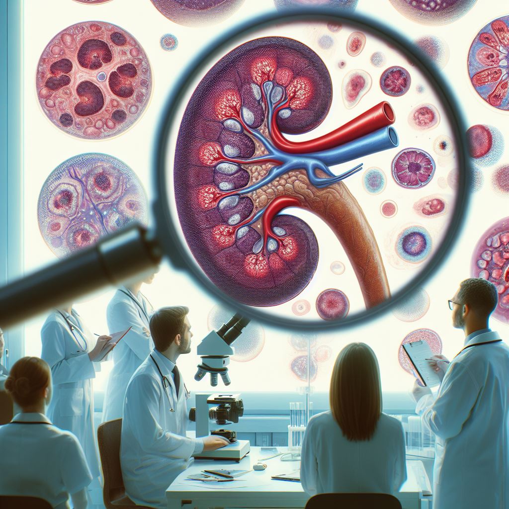
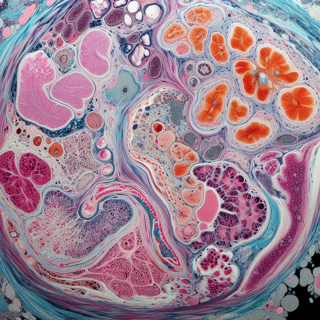
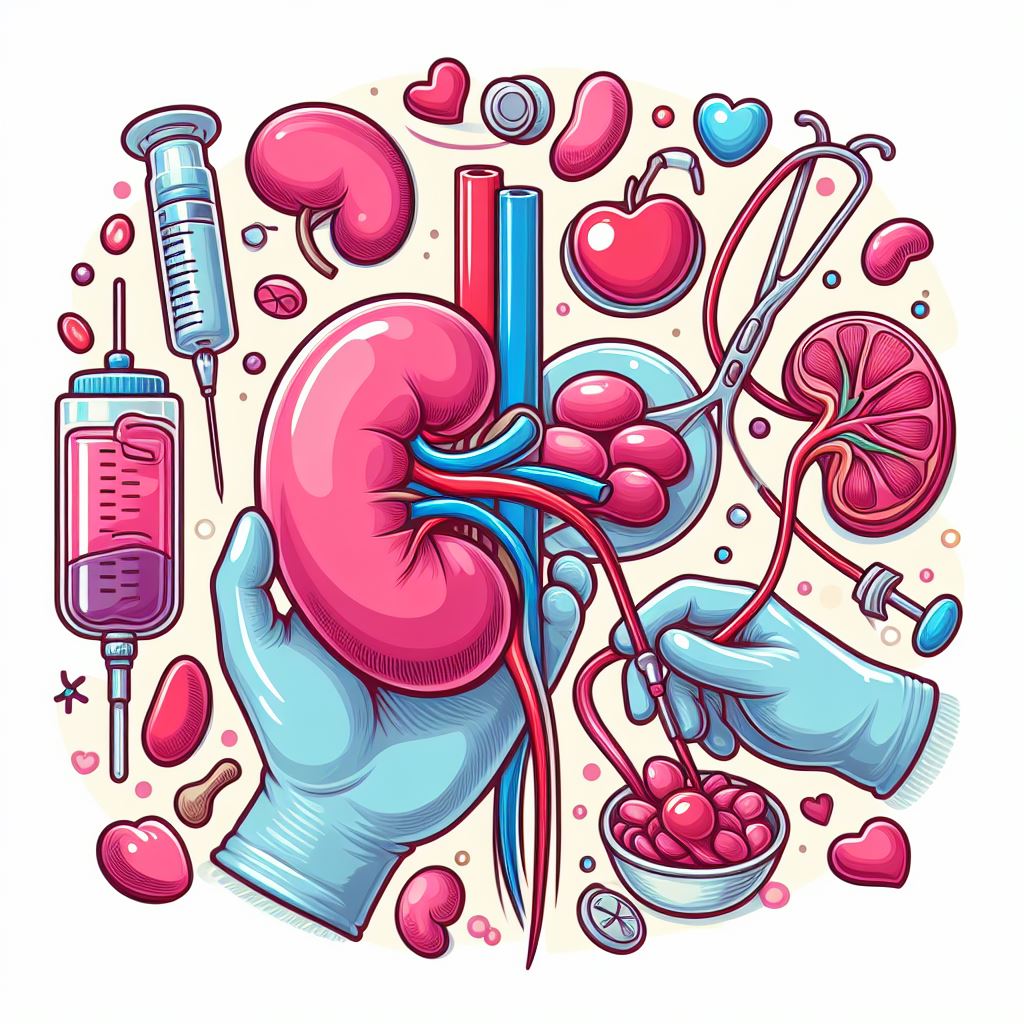
Kidney Biopsy Ilustration

Kidney Biopsy

A kidney biopsy, also known as renal biopsy or percutaneous kidney biopsy, is a diagnostic procedure that involves obtaining a small sample of kidney tissue for microscopic examination. This technique helps nephrologists and pathologists diagnose and evaluate various kidney disorders, assess the severity of the disease, and guide proper treatment strategies.

Indications for Kidney Biopsy. Kidney biopsy is typically performed when non-invasive diagnostic methods, such as blood tests, urine tests, or imaging studies, are inconclusive or insufficient for a definitive diagnosis. Common indications for kidney biopsy include:

  1. Unexplained acute kidney injury (AKI) or chronic kidney disease (CKD).
  2. Nephrotic syndrome or significant proteinuria (abnormal levels of protein in the urine).
  3. Haematuria (blood in the urine) of unknown origin.
  4. Glomerular diseases, such as glomerulonephritis or vasculitis.
  5. Monitoring kidney transplant rejection or assessing transplant dysfunction.
  6. Evaluating certain inherited kidney disorders.
  7. Diagnosing or staging kidney cancer.

Types of Kidney Biopsy. There are two primary types of kidney biopsy:

  1. Percutaneous kidney biopsy: This is the most common method, in which a needle is inserted through the skin to obtain a kidney tissue sample. The procedure is usually performed under local anaesthesia and imaging guidance, such as ultrasound or CT scan.
  2. Open kidney biopsy: This is a more invasive procedure, performed under general anaesthesia, where a surgeon makes an incision in the skin and directly visualizes the kidney to remove a tissue sample. Open kidney biopsy is less common and typically reserved for cases where percutaneous biopsy is contraindicated or has failed to yield adequate tissue.

The Kidney Biopsy Procedure. Before the biopsy, the patient may be instructed to stop taking certain medications, such as blood thinners, to minimize the risk of bleeding. The procedure generally involves the following steps:

  1. The patient lies face down or on their side, depending on the biopsy approach.
  2. The skin over the kidney is cleaned and sterilized, and local anaesthesia is administered to numb the area.
  3. The physician uses ultrasound or CT guidance to find the proper biopsy site and insert a special biopsy needle into the kidney.
  4. The needle is advanced into the kidney tissue, and a small sample is quickly extracted. The process may be repeated to obtain added samples if necessary.
  5. The needle is removed, and pressure is applied to the biopsy site to minimize bleeding. A bandage or dressing is applied to the area.
  6. The patient is watched for a few hours in a recovery area to ensure there are no immediate complications.

Risks and Complications. While kidney biopsy is generally considered safe, it carries some potential risks and complications, including:

  1. Bleeding: Minor bleeding at the biopsy site is common, but more severe bleeding may require blood transfusions or surgical intervention.
  2. Infection: There is a small risk of infection at the biopsy site or in the kidney.
  3. Injury to surrounding structures: Rarely, the needle may cause injury to nearby organs or blood vessels.
  4. Inadequate tissue sample: Occasionally, the biopsy may not supply enough tissue for a definitive diagnosis, causing a repeat procedure.

Benefits of Kidney Biopsy. Despite the risks, kidney biopsy offers significant benefits in the diagnosis and management of kidney-related conditions:

  1. Definitive diagnosis: A kidney biopsy can supply valuable information about the type and severity of kidney disease, helping guide right treatment and prognosis.
  2. Monitoring disease progression: In certain cases, kidney biopsies can help track the progression of kidney diseases and evaluate the effectiveness of treatment interventions.
  3. Personalized treatment: Biopsy results can help physicians tailor treatment plans to the specific needs of each patient, improving clinical outcomes.
  4. Assessing transplant rejection: For kidney transplant recipients, biopsies can help detect early signs of rejection, allowing for prompt intervention and improved transplant survival.

Post-Biopsy Care and Follow-Up. After the kidney biopsy, patients are typically advised to:

  1. Rest for 24-48 hours and avoid strenuous activities to minimize the risk of bleeding.
  2. Monitor the biopsy site for signs of infection, such as redness, swelling, or discharge.
  3. Report any unusual symptoms, like severe pain, fever, or persistent bleeding, to their healthcare provider.

Follow-up appointments are essential to review the biopsy results, discuss the diagnosis, and develop a treatment plan. The patient's healthcare team will supply personalized recommendations based on the findings and the individual's specific needs.

A kidney biopsy is an essential diagnostic tool that enables healthcare providers to obtain crucial information about kidney health and function. Though the procedure carries some risks, it plays a vital role in diagnosing various kidney disorders, guiding treatment decisions, and monitoring disease progression. With proper care and follow-up, a kidney biopsy can significantly contribute to improving patient outcomes and keeping kidney health.

Renal Picture Banner Renal Picture Banner Renal Picture Banner Renal Picture Banner Renal Picture Banner Renal Picture Banner Renal Picture Banner Renal Picture Banner Renal Picture Banner Renal Picture Banner Renal Picture Banner Renal Picture Banner Renal Picture Banner Renal Picture Banner Renal Picture Banner Renal Picture Banner Renal Picture Banner Renal Picture Banner Renal Picture Banner Renal Picture Banner
Interesting and helpful web links
Nephrological societies, organizations and associations
SNeS NTO STS ČNS ČTS PTN OEGfN OEGfN DGfN SIN ERA ASN ISN ISPD EuroPD CANS WKD
Associations and organizations of patients with kidney disease
SDAT National Kidney Federation
Health care facilities and educational institutions
PentaHospitals International PentaHospitals Nemocnica Bory PentaHospitals Slovensko
LF UK Bratislava UK Bratislava SZU LF UPJŠ Košice LF UPJŠ Košice UPJŠ Košice UN Bratislava Milosrdní bratia Ľubovnianska nemocnica, n.o.
Nephrology educational societies, organizations and associations
Medscape Healio UKIDNEY National Kidney Foundation KDIGO Medupdate EU
Nephrology Societies and Nursing Associations
EDTNA/ERCA ANNA
Slovakia - health insurance companies
Dôvera - zdravotná poisťovňa Všeobecná zdravotná poisťovňa Union zdravotná poisťovňa
Slovakia - state and self-governing institutions and bodies
SLOVENSKO.sk NCZI MZSR ÚDZS ÚVZSR eVÚC
Important European and global institutions
UN EU EP World Health Organisation
Proton NordVPN Surfshark Blinkist G2 Proton VPN Grammarly Skilmea
Audiolibrix www.martinus.sk

Websupport Websupport Websupport